What is Vertebral Stenting?
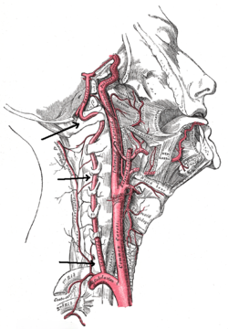
The vertebral arteries are vital blood vessels that supply blood to the brain, particularly the brainstem and cerebellum. When these arteries become narrowed or blocked due to atherosclerosis (plaque buildup) or other vascular conditions, it can significantly reduce blood flow and increase the risk of stroke or transient ischemic attacks (TIAs).
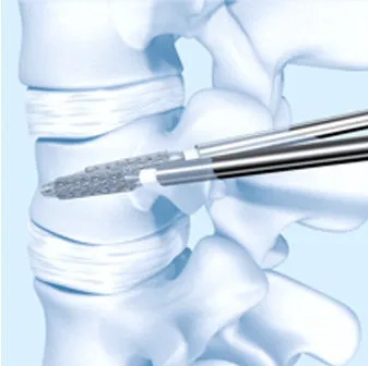
Vertebral stenting is a minimally invasive endovascular procedure that involves placing a small metallic mesh tube (stent) inside the narrowed vertebral artery. This helps widen the artery, restore proper blood flow, and reduce the risk of recurrent strokes. It is often performed in patients who do not respond well to medications alone.


Signs & Symptoms of Vertebral Artery Narrowing
Narrowing (stenosis) of the vertebral artery can cause symptoms related to reduced blood supply to the brain, such as:
-
Dizziness, imbalance, or unsteadiness while walking
-
Recurrent episodes of vertigo
-
Blurred or double vision
-
Difficulty speaking or slurred speech
-
Weakness or numbness in the face, arms, or legs
-
Sudden fainting episodes (drop attacks)
-
Transient ischemic attacks (mini-strokes)
-
Increased risk of posterior circulation stroke
If these symptoms are recurrent, evaluation by a neurologist is crucial.
Diagnostic Procedures
At the care of Dr. Apratim Chatterjee, diagnosis of vertebral artery narrowing involves:
Clinical Evaluation: Detailed history and neurological examination.
Doppler Ultrasound: Non-invasive test to assess blood flow in the neck vessels.
CT Angiography (CTA) / MR Angiography (MRA): High-resolution imaging to visualize arterial narrowing.
Digital Subtraction Angiography (DSA): Gold-standard test to confirm diagnosis and guide stent placement.
These investigations help determine the severity of narrowing and suitability for vertebral stenting.
These investigations help determine the severity of narrowing and suitability for vertebral stenting.


